Business Introduction
Introduction to Joint Device Center
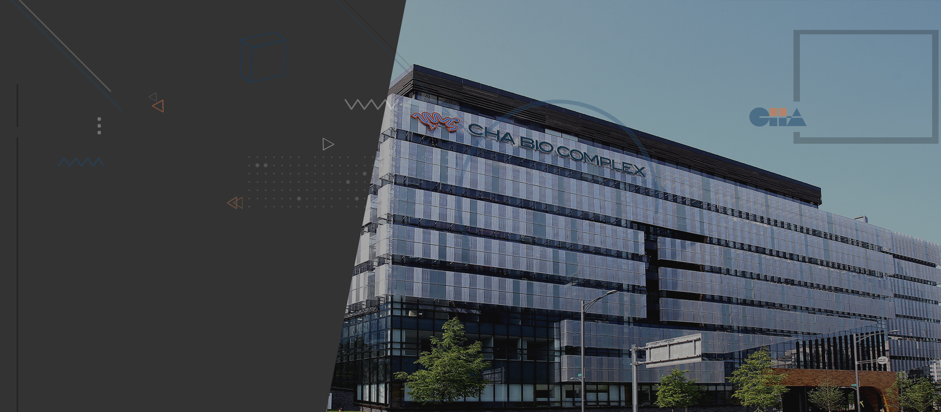
-On October 1, 2014, a research support project was carried out at the joint device center installed in the CHA Bio Complex.
– We operate a joint equipment room and tissue cell analysis room to open expensive equipment and support analysis research for the convenience of researchers.
Application and application procedures for use

* Inquiries: Administrative Office (031-881-7096) / Joint Equipment Room (031-881-7267) / Tissue Cell Analysis Room (031-881-7278)
* Joint Device Center Operation Guide and Precautions
– Device open to users: 24 hours a day, including weekends/nights, upon prior reservation for those who have completed device education and training (external users can use it during business hours (09:00-18:00))
– Education Inquiry: Please contact us at least 2 days in advance.
– Cancellation is possible on My Page. Please cancel at least 2 days in advance and contact the person in charge.
– For external users, please contact us in advance when reserving the device and enter your company name in the ‘Person in Charge of Research’ section.
Common equipment room available equipment and operating rates
Common equipment room equipment list
| no | Location | Device name (company/model name) | Purpose of use |
|---|---|---|---|
| 1 | Microscope room 1 | Confocal microscopy (Zeiss/LSM 880) | By identifying the three-dimensional structure of a fluorescently stained sample, there is less cross-talk when observing multi-fluorescently stained samples, and quantitative fluorescence analysis is possible. By optically sectioning colocalization samples, clear optical section images without blurring are possible. The shape and internal structure can be visualized without any damage to the sample, and the dedicated transmitted light detector provides high-resolution transmitted light images and is useful for analyzing the three-dimensional structure and micromorphology of samples such as chromosomes and cells. |
| 2 | Microscope room 3 | Confocal microscopy (Leica/TCS SP5II) | |
| 3 | Microscope room 2 | Laser Capture Microdissection (Arcturus LCM) | It is an essential equipment in molecular biology, cell biology, and pathology research enabling us to see related cells and tissues using a CCD camera, and then transfers and specialized cells within the tissue using a low-energy laser and special film. An automated system that easily obtains tissue by cutting it with a laser without causing chemical or physical changes in its shape or cells. |
| 4 | Microscope room 4 | Fluorescence microscope (Zeiss/LSM510) | A microscope that allows observation by fixing cells and staining them using a primary antibody labeled with a fluorescent substance, or by staining them with a secondary antibody labeled with a fluorescent substance when using an unlabeled primary antibody. |
| 5 | Microscope room 3 | Fluorescence microscope (Nikon/ECLIPSE Ni) | |
| 6 | All microscope rooms | Cryotome (Leica/CM3050S) | |
| 7 | All microscope rooms | Microtome (Leica/RM2125RTS) | |
| 8 | FACS room 2 | Slide Scanner (Zeiss/ AxioScan Z.1) | Equipment that creates more accurate and highly reproducible images by automating the task of scanning the entire slide with superior functionality and resolution than that of a general microscope using the digital slide method. |
| 9 | FACS room 1 | Flow Cytometry (BD/FACSCalibur) | This is a device that uses the characteristic of a fluorescent substance being activated and released by a laser beam to pass a fluorescently labeled liquid sample through a certain detection area and perform qualitative quantification by characteristic. By processing specific fluorescent compounds that can bind or bind to various organic substances within cells, the presence of specific components can be checked and quantitative information can be obtained. |
| 10 | FACS Room 1 | Flow Cytometry (Beckman/CytoFlex) | |
| 11 | FACS Room 3 | Flow Cytometry Sorter (Beckman) | |
| 12 | All FACS rooms | Ultrapure water production device | produces ultrapure water by electrical deionization, reverse osmosis, distillation, or deionization. |
| 13 | All FACS rooms | Autoclave (Tomy/SX-700) | The sterilization temperature ranges from 105℃ to 135℃ and the pressure is up to 40psig, which kills the sterilizer using steam and pressure. |
| 14 | FACS anterior chamber | Ultracentrifuge (Beckman/XL-90 ) | A method of separating cell organelles or macromolecules using centrifugal force generated by rotating the sample at ultra-high speed. Used for protein analysis using density gradient for separation of DNA or viruses. |
| 15 | FACS anterior chamber | Highcentrifuge (Beckman/Abanti J-E) | It is most often used to divide the homogenate into several parts using centrifugal force. The homogenate is placed in a test tube and the centrifuge is rotated at high speed to separate substances according to particle size and density. |
| 16 | FACS anterior chamber | Ice maker (POSIM/CM-50F) | |
| 17 | Basic equipment room 1 | Freeze dryer (Operon/FDS-7012) | The sample is placed in a container, pre-frozen at about -20°C or lower with dry ice and placed in a desiccator connected to a cold trap and vacuum pump. A heating plate supplies heat of sublimation for multiple freeze drying, and water vapor generated during drying is condensed into ice. Consists of a condenser, vacuum chamber, and vacuum pump. |
| 18 | IXM-Ctlf | High Contents Screeing Imagning System(MD/ImageXpress Micro Confocal ) | A microscopy instrument capable of switching between widefield and confocal imaging of fixed and live cells; an automated, high-content imaging system capable of observing thick tissues, 2D and 3D models, and intracellular structures; standard confocal mode (60 μm pinhole) (spinning disk with pinhole diameter), high-resolution confocal mode (spinning disk with pinhole diameter of 42 μm), etc. |
| 19 | Basic equipment room 2 | Luminescent image analyzer (GE/LAS 4000) | Luminescent image analyzer (GE/LAS 4000) An image obtained as a result of a blotting technique in which proteins separated and distributed on a gel are moved to a specific membrane in an electric field, and then only specific types of proteins are colored and detected using an antigen-antibody reaction. |
| 20 | Basic equipment room 2 | Luminometer (MD/SpectraMax-L) | A device that can detect and measure luminescence/changes in biological components. Expression measurement allows exploration of various biomarkers for environmental pollutants in vivo and is used to study changes in the structure of cell membranes and capable of tracking the movement of metal ions in the body. EROD ECOD and AHH expressed in cells and tissues. |
| 21 | Basic equipment room 2 | Luminex (Luminex/Luminex 200 system) | |
| 22 | Basic equipment room 2 | Microplate Reader (MD/Spectramax iD5) | Analyze interrelationships through experiments using materials with optical properties such as absorption, fluorescence, luminescence, temporal fluorescence (TRF), and fluorescence polarization. |
| 23 | Basic equipment room 2 | Gel-Doc. System (Syngeze/G:Box) | Used for DNA image analysis/western blotting on agarose gel after electrophoresis |
| 24 | Basic equipment room 2 | PCR machine (AB/Proflex Base) | An automatic device that artificially amplifies nucleic acids using polymerase chain reaction techniques. |
| 25 | Basic equipment room 2 | qRT-PCR machine (AB/ViiA7) | In real time PCR, PCR amplification products are monitored in real time and quantified in the area where amplification occurs exponentially. Therefore, unlike the existing RT-PCR method, accurate real-time PCR (polymerase chain reaction) is based on the amplification rate theory of PCR. It is a device that integrates a thermal cycler and a spectrofluorometer, and monitors the production process of PCR amplification products in real time to analyze the amount of target DNA. |
| 26 | Basic equipment room 2 | PXRD (bruker/D2 phaser) | is an X-ray diffraction analyzer suitable for crystal structure analysis of crystalline types using powder. |
| 27 | Basic equipment room 2 | hybridization oven (NBIOTECH/NB-202) |
Shared equipment room unit price details
| no | Device name (company/model name) | division | internal user | external user | notes |
|---|---|---|---|---|---|
| 1 | Confocal microscopy (Zeiss/LSM 880) | Imaging | 50,000 KRW/hour | 100,000 KRW/hour | When canceling a reservation, a fee will be charged. Cancellation the day before: 50%. Cancellation on the day: 70%. |
| 2 | Confocal microscopy (Leica/TCS SP5II) | Imaging | 20,000 KRW/hour | 40,000 KRW/hour | |
| 3 | Laser Capture Microdissection (Arcturus LCM) | Imaging | 20,000 KRW/hour | 40,000 KRW/hour | |
| 4 | Fluorescence microscope (Zeiss/LSM510) | Imaging | 5,000 KRW/hour | 10,000 KRW/hour | |
| 5 | Fluorescence microscope (Nikon/ECLIPSE Ni) | Imaging | 5,000 KRW/hour | 10,000 KRW/hour | |
| 6 | Cryotome (Leica/CM3050S) | – | 5,000 KRW/hour | 10,000 KRW/hour | |
| 7 | Microtome (Leica/RM2125RTS) | – | 3,000KRW/hour | 6,000 KRW/hour | |
| 8 | Slide Scanner (Zeiss/ AxioScan 2.1) | – | 30,000 KRW/hour | 60,000 KRW/hour | |
| analyze | free | free | |||
| 9 | Flow Cytometry (BD/FACSCalibur) | Analysis | 30,000 KRW/hour | 60,000 KRW/hour | Broken |
| 10 | Flow Cytometry (Beckman/CytoFlex) | Analysis | 30,000 KRW/hour | 60,000 KRW/hour | |
| 11 | Flow Cytometry Sorter (Beckman) | Bundang Cha Hospital Inquiry | |||
| 12 | Ultrapure water manufacturing equipment | – | 12,000 KRW/10L | 24,000
KRW /10L |
|
| 13 | Autoclave (Tomy/SX-700) | – | 3,000 KRW/hour | 6,000 KRW/hour | |
| 14 | Ultracentrifuge (Beckman/XL-90 ) | – | 10,000 KRW/hour | 20,000 KRW/hour | Can be used up to 24000rpm |
| 15 | Highcentrifuge (Beckman/Abanti J-E) | – | 10,000 KRW/hour | 20,000 KRW/hour | |
| 16 | Ice maker (POSIM/CM-50F) | – | free | free | |
| 17 | Freeze dryer (Operon/FDS-7012) | – | 10,000 KRW/day | 20,000 KRW/day | |
| 18 | High Contents Screeing Imagning System(MD/ImageXpress Micro Confocal ) | – | 40,000 KRW/day | 80,000 KRW/day | |
| analyze | free | free | |||
| 19 | Luminescent image analyzer (GE/LAS 4000) | – | free | free | Open from November 2018 |
| 20 | Luminometer (MD/SpectraMax-L) | – | 2,000 KRW/hour | 4,000 KRW/hour | At least 30 minutes / per session |
| 21 | Luminex (Luminex/Luminex 200 system) | – | 5,000 KRW/hour | 10,000 KRW/hour | At least 30 minutes / per session |
| 22 | Microplate Reader (MD/Spectramax iD5) | – | 6,000 KRW/hour | 12,000 KRW/hour | At least 30 minutes / per session |
| 23 | Gel-Doc. System (Syngeze/G:Box) | – | 2,000 KRW/hour | 4,000KRW/hour | At least 30 minutes / per session |
| 24 | PCR machine (AB/Proflex Base) | – | 2,000KRW/hour | 4,000 KRW/hour | At least 30 minutes / per session |
| 25 | qRT-PCR machine (AB/ViiA7) | – | 5,000 KRW/hour | 10,000 KRW/hour | At least 30 minutes / per session |
| 26 | PXRD (bruker/D2 phaser) | – | 10,000 KRW/hour | 20,000 KRW/hour | |
| 27 | hybridization oven (NBIOTECH/NB-202) | – | free | free | |
| * | virus lab a | – | 10,000KRW/day | 20,000 KRW/day | CO2 Incubator, Clean Bench, Shaking Incubator, etc. |
| * | virus lab b | – | 200,000 KRW/month | 400,000 KRW/month | CO2 Incubator, Clean Bench, Shaking Incubator, etc. |
Tissue Analysis Laboratory Tissue Analysis Operation Rate
조직세포분석실 실험 단가내역
| item | Unit price (KRW) | item | Unit price (KRW) | |||
| A | Preprocessing and breakdown | 4,000 | E | immunohistochemistry | ||
| B | Block production | single stain | 50,000 | |||
| paraffin(Manual, Automatic) | 9,000 | F | immunofluorescence | |||
| cryo | 5,000 | single stain | 45,000 | |||
| C | section | double stain | 50,000 | |||
| Section for dyeing (Paraffin) | 3,500 | G | TUNEL stain | |||
| Section for dyeing (Cryo) | 4,000 | Basic size (less than 0.5cm) | 50,000 | |||
| Unstain slide 용 | 4,000 | Large size (over 0.5cm) | 70,000 | |||
| Whole section | 4,500 | H | Analysis (*Available after consultation) | |||
| interval section | 4,000 | Carl Zeiss Slide Scanner | Normal | 30,000 | ||
| D | staining | Neon | 40,000 | |||
| H&E | 5,000 | Organize PowerPoint results | 100,000~ | |||
| PAS | 10,000 | G | External and company-affiliated samples | 30% of the unit price will be charged separately from 21.07.01 onwards. | ||
| Alcian blue | 10,000 | |||||
| Masson’s Trichrom | 10,000 | |||||
| Oil red O | 15,000 | |||||
| Other dyeing | 10,000~ | |||||
조직세포분석실 기기사용 단가
| 기기명 | 수량 | Model | 회사 | 단가 |
|---|---|---|---|---|
| Cryotome | 1 | CM3050 | Leica | 1000/1hr |
| Microtome | 1 | RM2125RT | Leica | 1000/1hr |
| Tissue warmer | 2 | 26007CE | Barnstend | 1000/1hr |
| microscope | 1 | Nikon | 2000/1hr | |
| 타사 연구기관에서 사용할 경우 사용 단가는 기존 단가의 *3 | ||||
기기 소모품 사용 단가 내역
| 소모품 | 단가 |
|---|---|
| Cryotome blade | 3000원/장 |
| Microtome blade | 3000원/장 |
| O.C.T Compound | 200원/sample |
| coating slide glass | 1000원/장 |
| paraplast | 2000원/sample |
| Embedding cassette | 100원/sample |
| Slide box | 6000원/개 |